Figure 1 from Brain surface temperature under a craniotomy.
Por um escritor misterioso
Last updated 16 abril 2025

Fig. 1. Rapid cooling of the brain surface in an in vivo mouse preparation. A: schematic representation of a cranial window during recording of temperature and single-cell activity in the anesthetized mouse. The main potential routes of heat transfer are indicated. B: brain surface temperature measured with the thermocouple during replacement of the artificial cerebrospinal fluid (ACSF) with fresh ACSF warmed to 38°C. ACSF was replaced twice, indicated by the arrowheads. - "Brain surface temperature under a craniotomy."

Astrocyte-neuron lactate shuttle plays a pivotal role in sensory-based neuroprotection in a rat model of permanent middle cerebral artery occlusion
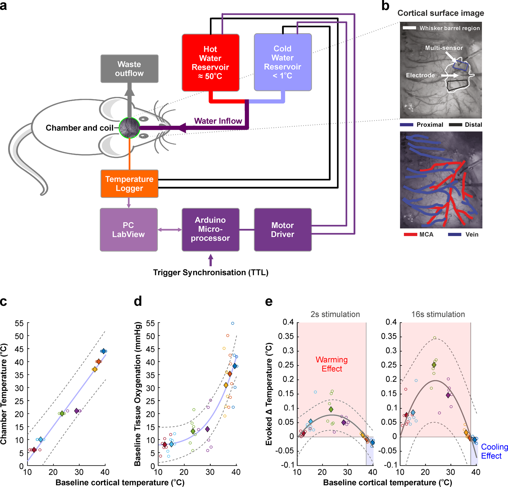
Bidirectional alterations in brain temperature profoundly modulate spatiotemporal neurovascular responses in-vivo

Photothrombotic Middle Cerebral Artery Occlusion in Mice: A Novel Model of Ischemic Stroke
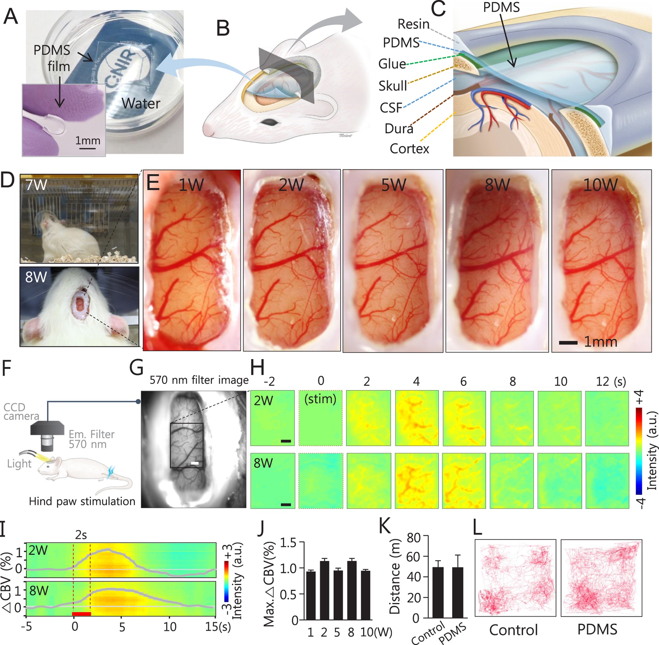
A soft, transparent, freely accessible cranial window for chronic imaging and electrophysiology
Assessment of Thermal Damage from Robot-Drilled Craniotomy for Cranial Window Surgery in Mice

Brain surface temperature under a craniotomy

Transient temperature changes at the brain surface during 15k RPM

Figure 1 from Brain surface temperature under a craniotomy.

Maximum temperature recorded during drilling of rat craniotomy.

Regional temperature and quantitative cerebral blood flow responses to cortical spreading depolarization in the rat - Chunyan Li, Raj K Narayan, Ping Wang, Jed A Hartings, 2017

Data collection and craniotomy. Left: The infrared camera setup is
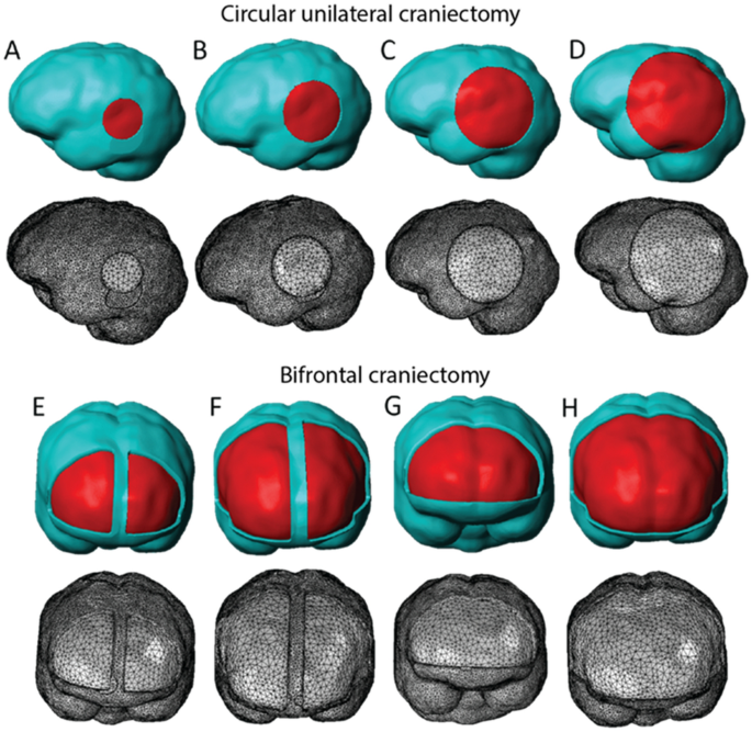
Decompressive craniectomy of post-traumatic brain injury: an in silico modelling approach for intracranial hypertension management

Craniotomy for acute monitoring of pial vessels in the rodent brain - ScienceDirect

Longitudinal two-photon calcium imaging with ultra-large cranial window for head-fixed mice - ScienceDirect
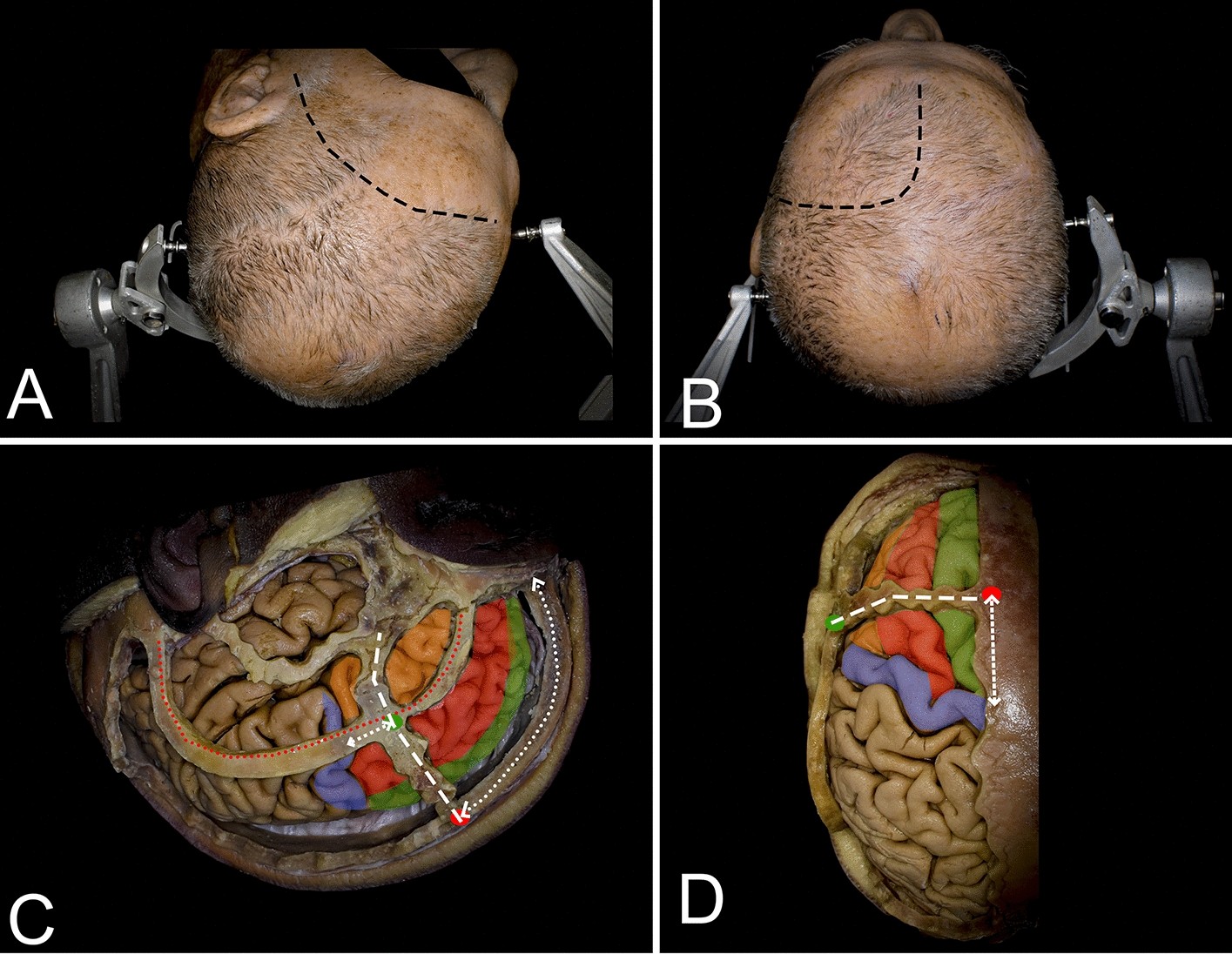
Cortical and white matter anatomy relevant for the lateral and superior approaches to resect intraaxial lesions within the frontal lobe
Recomendado para você
-
 BRAIN TEST NÍVEL 367 EM PORTUGUÊS16 abril 2025
 -
 Brain Test Nivel 367 - Quiere tener grandes músculos16 abril 2025
 -
 Brain Test, Nivel 367, Quiere tener grandes musculos, Explicado Español16 abril 2025
 -
 Association of body mass index and waist-to-hip ratio with brain structure16 abril 2025
 -
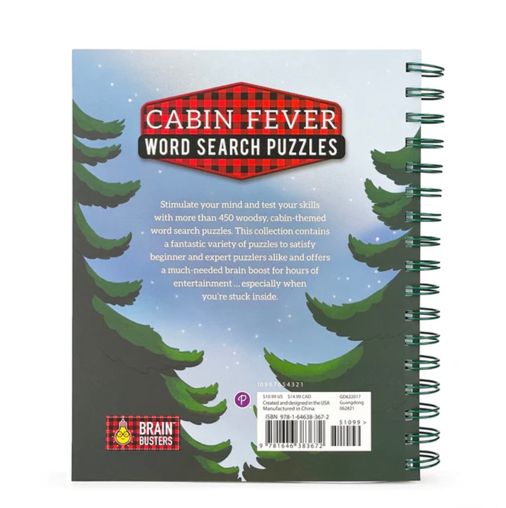 Cabin Fever Word Search Puzzle – General Store of Minnetonka16 abril 2025
 -
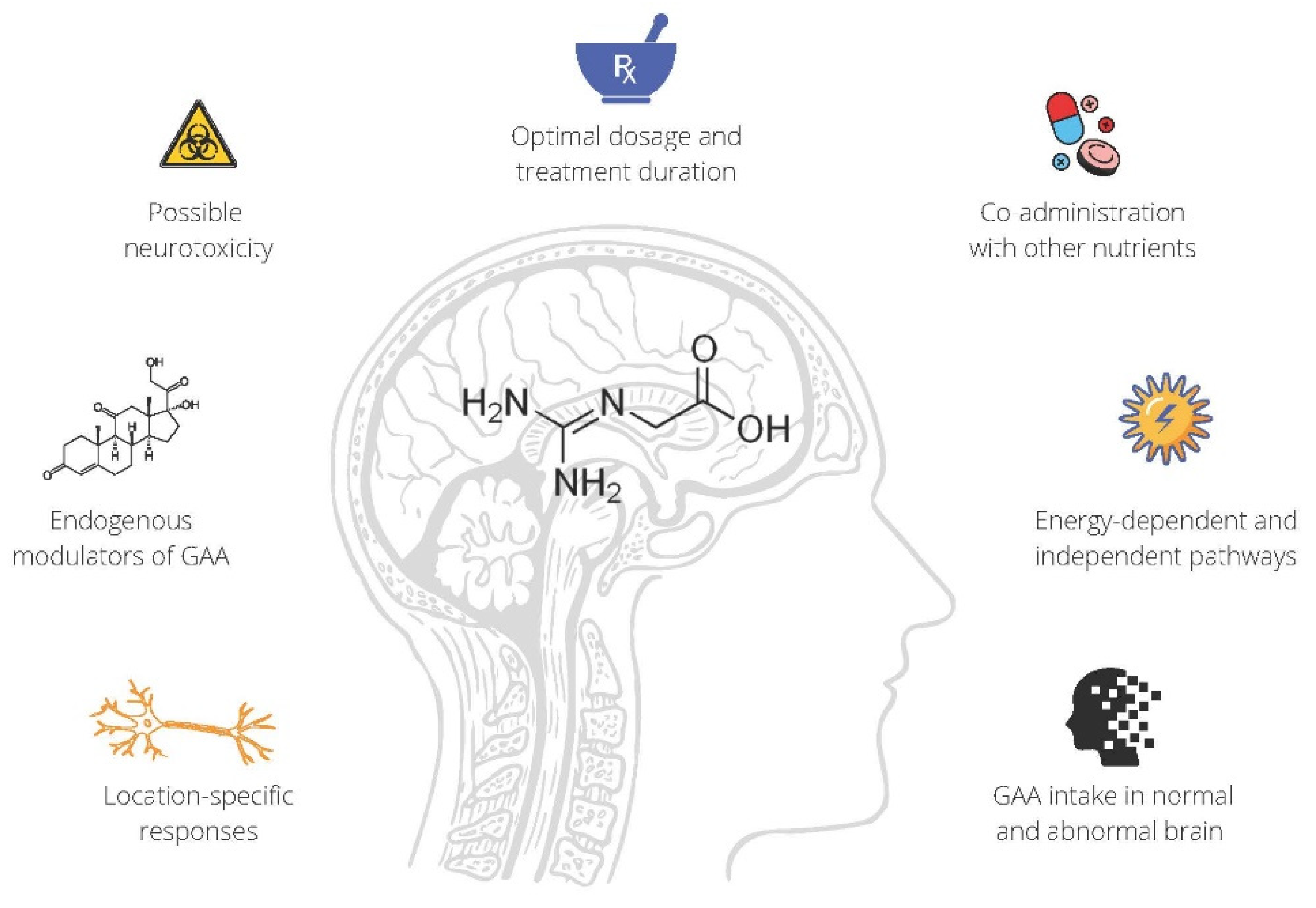 Nutrients, Free Full-Text16 abril 2025
 -
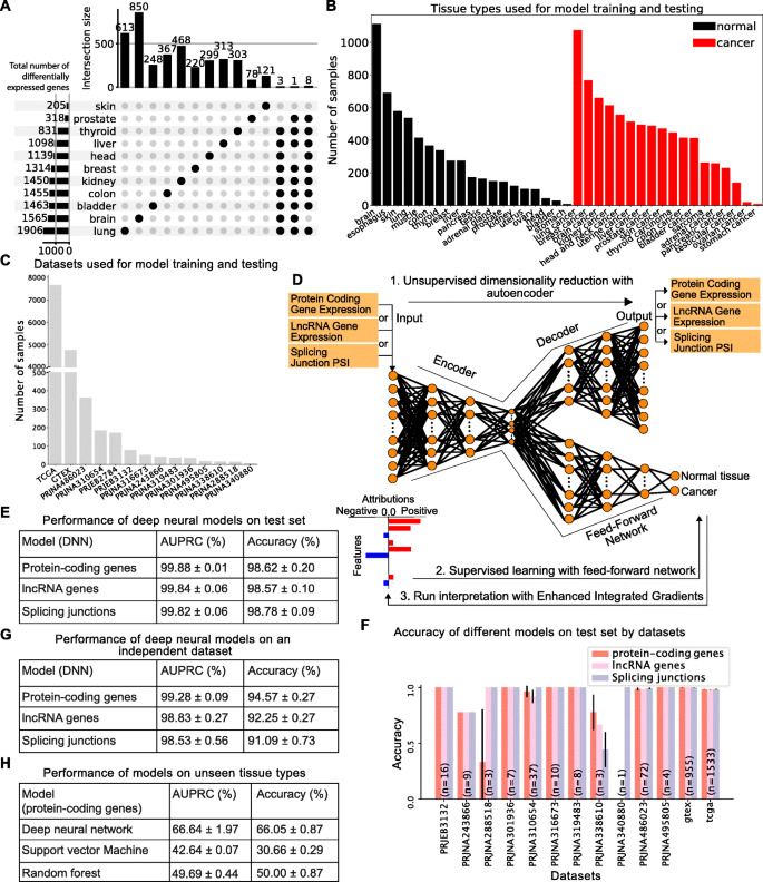 Identifying common transcriptome signatures of cancer by interpreting deep learning models, Genome Biology16 abril 2025
 -
 Fluid transport in the brain16 abril 2025
 -
 Effectiveness of management strategies for uninvestigated dyspepsia: systematic review and network meta-analysis16 abril 2025
 -
 Pharmacology, Biochemistry and Behavior 1989: Vol 33 Index : Free Download, Borrow, and Streaming : Internet Archive16 abril 2025
você pode gostar
-
 How to Install NOOBs16 abril 2025
 -
man face vs woman face roblox|TikTok Search16 abril 2025
-
 10 filmes de caçadores de tesouros parecidos com Indiana Jones16 abril 2025
 -
 Where are all the forks? - Side-Eyes Chloe16 abril 2025
 -
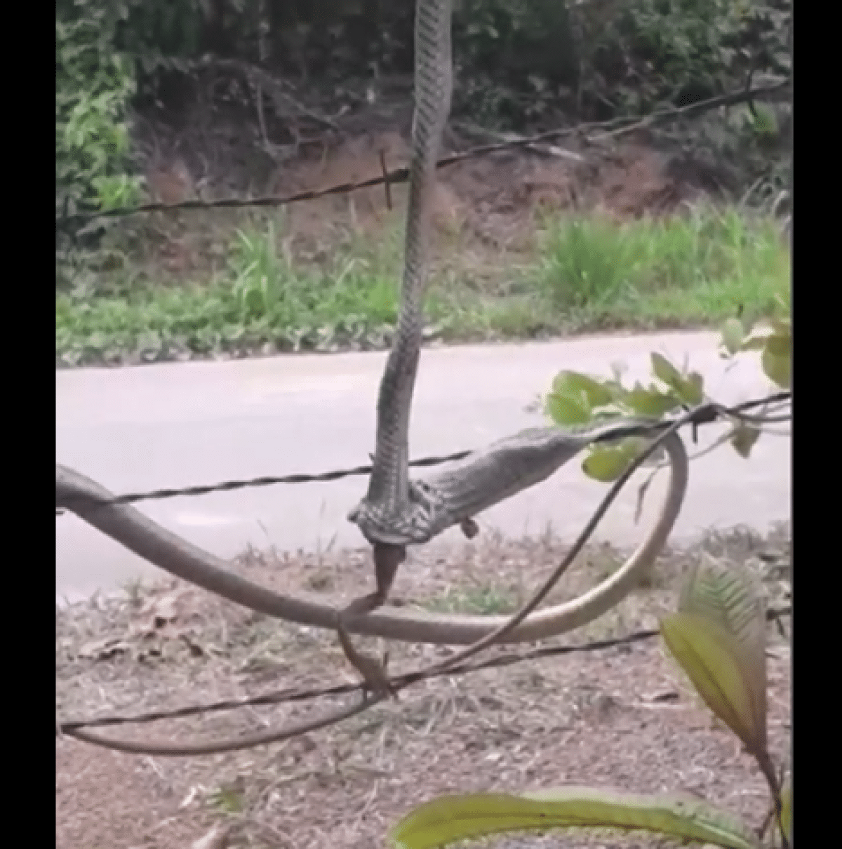 Veja a disputa intrigante entre duas cobras para devorar uma perereca - CenárioMT16 abril 2025
 -
![DISC] The Breaker 3 – Eternal Force - Chapter 86 : r/TheBreaker](https://external-preview.redd.it/disc-the-breaker-3-eternal-force-chapter-86-v0-LkPU__azqHMnwrmjft8k72UFi1xtRHegmZ2rL4XKv-A.jpg?auto=webp&s=a51d50e63e1c6fc367fedb0586d96f3a25f81607) DISC] The Breaker 3 – Eternal Force - Chapter 86 : r/TheBreaker16 abril 2025
DISC] The Breaker 3 – Eternal Force - Chapter 86 : r/TheBreaker16 abril 2025 -
 BORUTO TWO BLUE VORTEX16 abril 2025
 -
Mods for Alphabet Lore Melon - Apps on Google Play16 abril 2025
-
 GABINETE GAMER GAMEMAX KREATOR, RGB, LATERAL VIDRO TEMPERADO, S/ FAN, S/ FONTE, PRETO - KREATOR - GABINETES - Gamemax16 abril 2025
 -
 Life cycles woman stages growing up from Vector Image16 abril 2025